- Graduate Field Affiliations
- Biomedical Engineering
- Biomedical and Biological Sciences
- Mechanical Engineering
Biography
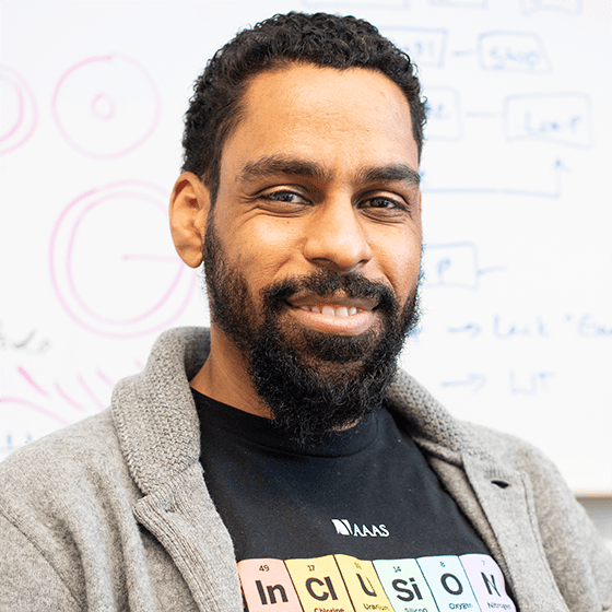
Dr. Karl Lewis joined the Meinig School as an assistant professor in July 2020. Dr. Lewis’s research interests center on understanding the interplay of mechanical cues and biological changes in musculoskeletal tissues. As a trained engineer, Dr. Lewis views the body as a system of sub-systems with a wide range of input and output signals. Cytokines, neurotransmitters, and hormones are some of the signals available for the body to use. Mechanical forces can modulate these signals and are critical for the health of musculoskeletal cells.
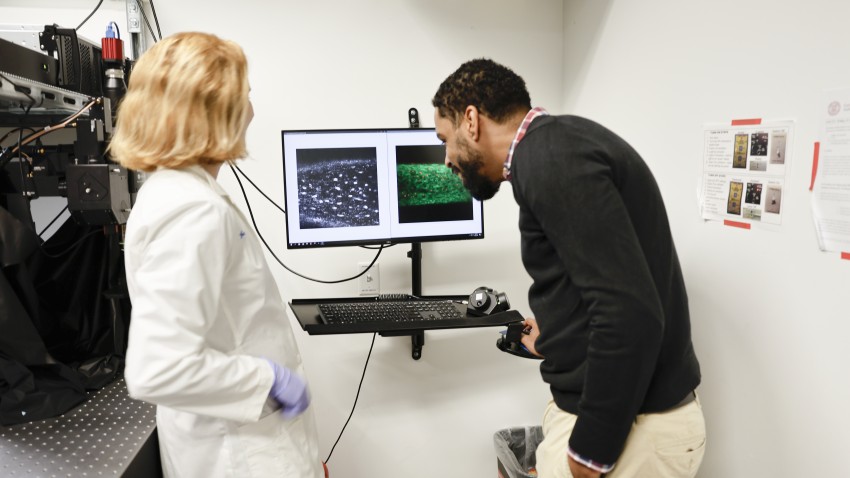
With this in mind, Dr. Lewis’s work seeks to understand how the acute sensing mechanisms in musculoskeletal cells relate to tissue-level changes in healthy and disease states. To interrogate these questions, Dr. Lewis works with cutting-edge intravital imaging techniques to observe cell signaling in real time. Using genetically modified mice, he and his collaborators are searching for proteins related to bone growth, development, and disease (i.e. osteoporosis).
Dr. Lewis comes to Cornell most recently from Indiana University School of Medicine, where he worked on two projects with Dr. Alex Robling as a post-doctoral fellow in the Department of Anatomy and Cell Biology. One project investigated the role of neurotransmitters in osteocyte mechanobiology using genetically modified mice bred in-house. The second project utilized intravital fluorescent imaging techniques to observe changes in osteocyte calcium signaling a function of genetic modification or pharmacological intervention.
At Cornell, the Lewis laboratory will continue this research in bone and also develop new intravital imaging techniques for studying mechanotransduction in other musculoskeletal tissues. The lab will interrogate how the acute sensing mechanisms in musculoskeletal cells relate to tissue-level changes in healthy and disease states, with the objective to use this knowledge to identify new targets for therapeutic intervention in musculoskeletal disease. Bone investigations will focus on understanding how bone cells interpret mechanical loading in real time using intravital imaging techniques. The lab will also work to understand how bone interacts with other neighboring tissues in the onset and progression of diseases like osteoarthritis and osteoporosis.
Research Interests
- Biomechanics and Mechanobiology
- Biomedical Engineering
- Molecular and Cellular Engineering
- Image Analysis
- Biomedical Imaging and Instrumentation
- Bioengineering
Select Publications
-
KJ Lewis, RB-J Choi, EZ Pemberton, WA Bullock, AB Firulli, AG Robling, “Twist1 inactivation in Dmp1-expressing cells increases bone mass but does not affect the anabolic response to sclerostin neutralization,” Int. J. Mol. Sci (2019)
-
KJ Lewis, WR Thompson, AG Robling, “The mTORC2 component Rictor is required for development of osteocyte cell processes and mechanical signaling,” JBMR (Submitted)
-
WA Bullock, AM Hoggatt, DJ Horan, KJ Lewis, H Yokota, S Hann, ML Warman, A Sebastian, GG Loots, FM Pavalko, AG Robling, “Expression of a Degradation‐Resistant β‐Catenin Mutant in Osteocytes Protects the Skeleton From Mechanodeprivation‐Induced Bone Wasting,” JBMR (Published, Early View 2019)
-
KJ Lewis, D Frikha-Benayed, J Louie, S Stephen, DC Spray, MM Thi, Z Seref-Ferlengez, RJ Majeska, S Weinbaum, MB Schaffler, “Osteocyte calcium signals encode strain magnitude and loading frequency in vivo,” PNAS 114(44):11775-11780 (2017)
-
C McGoverin, K Lewis, X Yang, M Bostrom, and N Pleshko, “The Contribution of Bone and Cartilage to the Near-Infrared Spectrum of Osteochondral Tissue,” Appl. Spectrosc. 68, 1168-1175 (2014)
Select Awards and Honors
- IFMRS Travel Grant for European Calcified Tissue Society Congress 2019
- NIH T-32 Training Grant, National Institute of Health 2017-2019
- Wallace H. Coulter Award for Academic Service 2015
- NASA New York Space Consortium Fellowship 2013-2017
- Alfred P. Sloan Graduate Scholarship for Minority Ph.D.s 2011
Education
- B.S., Mechanical Engineering (Biomedical Engineering Concentration), Temple University 2011
- Ph.D., Biomedical Engineering, Osteocyte Mechanobiology, The City College of New York 2017
- Postdoc (Osteocyte Mechanobiology, Bone Mechanotransduction), Indiana University School of Medicine 2020


